Total breakdown in the brain

What happens when nerve cells stop working?
A stroke is just one example of a condition when communication between nerve cells breaks down. Micro-failures in brain functioning also occur in conditions such as depression and dementia. In most cases, the lost capacity will return after a while. However, consequential damage will often remain so that the functional capability can only be restored through lengthy treatment — if at all. For this reason, researchers at the Chair of Psychiatry and Psychotherapy have been investigating what happens during such breakdown phases and looking at possible ways of preventing damage and speeding up the healing processes. Their findings have been recently published in the eminent journal Scientific Reports.
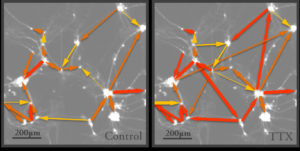
The research team headed by Jana Wrosch of the Chair of Psychiatry and Psychotherapy found that significant alterations occurred in neural cells while the communication pathways were blocked. Neuron networks reconnect during such periods of inactivity and become hypersensitive. If we imagine that normal communication pathways are motorways, when they are blocked a form of traffic chaos occurs in the brain whereby information is re-routed in disorganised form along what can be called side streets and minor routes. Additional synapses are generated everywhere and begin operating. When the signal is reinstated, the previously coordinated information routes no longer exist and, as in the case of a child, the appropriate functions need to be learned from scratch. Since they are receiving no normal signals during the phase of brain malfunction, the nerve cells also become more sensitive in an attempt to find the missing input. Once the signals return, this means they may overreact.
Nerve cells flicker when stained
Visualising the microscopically minute connections between the nerve cells is a major technical challenge. The conventional microscopic techniques currently available, such as electron microscopy, always require preliminary treatment of the nerve cells that are to undergo examination. However, this causes the nerve cells to die, so that the alterations that occur in the cells cannot be observed. To get round this problem, J. Wrosch and her team have developed a high-speed microscopy process along with special statistical computer software that make it possible to visualise the communication networks of living neurons. First, a video of the cells is made whereby an image is taken every 36 milliseconds. A special dye is used to stain the cells to ensure that the individual cells flicker whenever they receive a signal. Subsequently, the software recognizes these cells on the video images and detects the information pathways by which the signals are transmitted from cell to cell.
The nerve cells are then exposed to the pufferfish poison tetrodotoxin to simulate the blocking of communication channels that occurs in disorders. After inducing communication breakdown phases of varying lengths, the researchers remove the toxin from the cells and determine how the nerve cell networks have changed during exposure.
Thanks to this concept, we have been finally able to discover what happens when communication is blocked. Now we can try to develop medications that will help prevent these damaging changes.
explains J. Wrosch. In future projects, the research team plans to examine the exact mode of action of anti-depressants on nerve cell networks and intends to find new approaches to creating more effective drugs.
Further information
Jana Katharina Wrosch
Phone: +49 9131 8544294